
Phase Contrast Microscopy in Dentistry Isn’t New. It’s Finally Gaining the Attention It Deserves.
When dental professionals first see phase contrast microscopy being used chairside, they sometimes assume it’s a new trend.
It isn’t.
The use of live microscopy to study periodontal biofilm dates back nearly 50 years. What’s new is the momentum it’s gaining as more dental teams recognize the value of showing patients the living microbial picture and combining it with bacterial DNA-PCR testing.
Microscopy helps us see what’s happening in the biofilm. DNA-PCR testing helps us identify which specific bacteria are present.
They’re showing us different things, which is exactly why I believe they’re so powerful when used together.
The Scientific Roots Go Back to the 1970s
In 1978, Drs. Paul Keyes, Walter Wright, and Sigmund Howard published a report describing the use of phase contrast microscopy in the diagnosis and treatment of periodontal lesions. Their work used microscopy to identify microbial changes and monitor the response to therapy. Read the Keyes, Wright, and Howard paper. (https://pubmed.ncbi.nlm.nih.gov/351678/)
That same year, Drs. Max Listgarten and Lars Helldén used darkfield microscopy to compare bacteria collected from clinically healthy and periodontally diseased sites. They documented differences in the bacterial shapes, or morphotypes, associated with health and disease. Read the Listgarten and Helldén study. (https://pubmed.ncbi.nlm.nih.gov/350909/)
Darkfield and phase contrast aren’t identical technologies, but both allow us to examine live, unstained microorganisms in a wet mount. These early studies established an important concept: the microbial community associated with periodontal health doesn’t look the same as the community found in periodontal disease.
This didn’t begin with social media or a new microscope company. It began in periodontal research almost 50 years ago.
What Can We See With Phase Contrast Microscopy?
Most live oral microorganisms are nearly transparent on an unstained slide. Their refractive properties are similar to the fluid surrounding them, which makes them difficult to see with an ordinary brightfield microscope.
Phase contrast microscopy turns those subtle differences into visible contrast. It allows us to observe:
• The overall density of the biofilm
• Cocci, rods, filaments, and spirochete-shaped organisms
• The amount and type of microbial movement
• White blood cells and other host cells
• Organisms that appear consistent with amoebae or trichomonads
• Changes in the microbial picture as treatment progresses
The shapes matter, but they have to be interpreted correctly. A microscope can’t tell us that every round bacterium is beneficial or that every rod is pathogenic. It also can’t reliably identify most bacteria to the species level based on appearance alone.
What it can show us is the overall microbial pattern. A biofilm dominated by highly motile organisms and spirochetes isn’t the microbial picture typically associated with periodontal health. A less motile community dominated by coccoid forms is more commonly associated with health or improvement following treatment.
What Does the Microscopy Research Show?
• 1982: Researchers used darkfield microscopy to monitor subgingival bacterial changes during periodontal therapy. This demonstrated how microbial morphology and motility could be followed as treatment progressed. Read the periodontal monitoring study. (https://pubmed.ncbi.nlm.nih.gov/6757407/)
• 1982: Listgarten and Schifter evaluated differential darkfield microscopy as an aid in establishing individualized periodontal recall intervals. Read the recall interval study. (https://pubmed.ncbi.nlm.nih.gov/6964678/)
• 1986: An experimental gingivitis study found relatively high proportions of spirochetes with phase contrast microscopy after gingival inflammation was induced. Read the experimental gingivitis study. (https://pubmed.ncbi.nlm.nih.gov/3462202/)
• 1988: Trope, Tronstad, Rosenberg, and Listgarten used darkfield microscopy to help differentiate periodontal abscesses from endodontic abscesses. Periodontal samples contained substantially higher percentages of spirochetes. Read the abscess differentiation study. (https://pubmed.ncbi.nlm.nih.gov/3162933/)
• 1998: Researchers examined 200 diseased periodontal sites and 44 healthier control sites. Spirochetes were found in 197 of the 200 deep periodontal pockets, compared with 19 of the 44 control sites. Molecular testing also revealed that many types of treponemes can look similar under the microscope. Read the oral treponeme study. (https://journals.asm.org/doi/10.1128/jcm.36.5.1399-1403.1998)
• 2022: A study involving 60 patients with moderate periodontitis incorporated phase contrast microbial analysis into a periodontal maintenance protocol. Read the 2022 study. (https://www.mdpi.com/2411-5134/7/4/104)
• 2024: A report involving 20 patients used phase contrast microscopy at repeated visits to evaluate periodontal biofilm during treatment. Investigators tracked motile bacteria, spirochetes, inflammatory cells, and organisms interpreted as Entamoeba gingivalis, along with bleeding and pocket depths. The results were encouraging, although larger controlled studies are still needed. Read the 2024 microscopy report. (https://pmc.ncbi.nlm.nih.gov/articles/PMC11797751/)
This research doesn’t mean microscopy can diagnose every periodontal condition on its own. Some older studies found that live microscopy couldn’t reliably separate gingivitis from periodontitis without additional clinical information.
That doesn’t make the microscope less valuable. It tells us how it should be used.
Phase contrast microscopy is an adjunct to the comprehensive periodontal assessment. It adds a living, real-time view of the biofilm to the information we’re already collecting.
The Microscope Makes the Invisible Visible
This is where I believe microscopy becomes especially powerful in patient care.
We can talk to patients about bacteria, inflammation, bleeding, and infection, but they can’t see what’s living below their gumline. The microscope gives us a close-up view of the patient’s own biofilm.
They’re not looking at a generic photograph from a textbook or a scary image from the internet. They’re looking at a sample collected from their own mouth.
That visual can help patients understand why their gums are bleeding, why treatment has been recommended, why home care needs to change, and how the microbial picture can change following treatment.
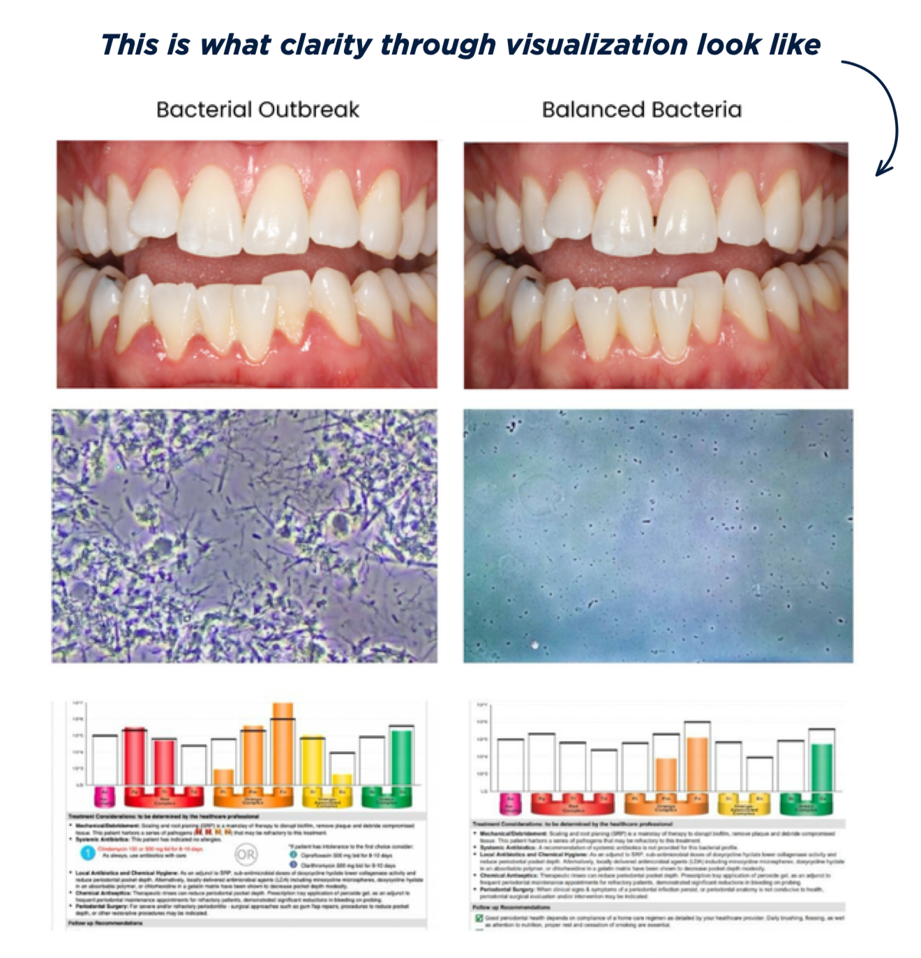
Microscopy transforms an abstract conversation into something patients can see. It allows us to show instead of simply tell.
But the image is only one part of the story.
Why We Need DNA-PCR Testing Too
The microscope gives us the living picture, but it can’t reliably identify most bacteria by species.
A spirochete moving under the microscope can’t automatically be labeled Treponema denticola. A small rod can’t be visually identified as Porphyromonas gingivalis. Important periodontal pathogens may also be present at levels that aren’t easily recognized microscopically.
That’s where bacterial DNA-PCR testing becomes so valuable.
Depending on the laboratory and collection method, the sample may come from saliva, an oral rinse, or subgingival plaque collected with paper points. PCR technology can identify and quantify specific bacterial targets, including:
• Porphyromonas gingivalis or Pg
• Aggregatibacter actinomycetemcomitans or Aa
• Treponema denticola
• Tannerella forsythia
• Fusobacterium nucleatum
• Prevotella intermedia
In 2007, researchers compared subgingival samples with oral rinse samples using real-time PCR. The testing detected and quantified major periodontal pathogens, including Pg and Aa. The authors concluded that oral rinse PCR could simplify microbial testing and potentially help monitor periodontal treatment. Read the oral rinse PCR study. (https://pubmed.ncbi.nlm.nih.gov/17199543/)
In 2009, investigators developed a quantitative PCR test for five major periodontal pathogens. In 165 saliva samples, the combined pathogen burden helped distinguish subjects with periodontitis from those without it. Read the five-pathogen PCR study. (https://pubmed.ncbi.nlm.nih.gov/19586997/)
A 2015 study also evaluated salivary levels of four major periodontal pathogens and found that the combined bacterial burden provided useful diagnostic information. Read the salivary pathogen burden study. (https://pubmed.ncbi.nlm.nih.gov/26484315/)
PCR testing gives us species-specific information that we can’t get from morphology alone. However, a positive PCR result doesn’t diagnose periodontal disease by itself. The results still have to be interpreted alongside bleeding, probing depths, clinical attachment levels, radiographic bone loss, medical history, and other clinical findings.
What About Oral Parasites?
Phase contrast microscopy can reveal larger motile organisms that may appear consistent with oral protozoa, including Entamoeba gingivalis and Trichomonas tenax.
A 2014 study used PCR to detect E. gingivalis in diseased periodontal pockets while finding it largely absent from healthy sites. Read the Entamoeba gingivalis PCR study. (https://pmc.ncbi.nlm.nih.gov/articles/PMC4077299/)
A 2021 study evaluating both E. gingivalis and T. tenax also found greater colonization among patients with periodontal disease, although the organisms weren’t completely absent from healthy participants. Read the oral protozoa study. (https://pmc.ncbi.nlm.nih.gov/articles/PMC8688919/)
Their exact role in periodontal disease is still being studied. An organism may look consistent with an amoeba or trichomonad under phase contrast, but microscopy alone shouldn’t be presented as definitive species confirmation. PCR is more specific when the appropriate protozoal targets are included in the test.
The Microscope and DNA-PCR Testing Answer Different Questions
Phase contrast microscopy helps us ask:
• What does the living biofilm look like right now?
• Are there high levels of motility, visible spirochetes, or inflammatory cells?
• How does this site’s microbial picture appear to change during care?
DNA-PCR testing helps us ask:
• Which targeted bacterial species were detected?
• Are Pg, Aa, Td, Tf, or other tested bacteria present?
• What’s the reported level of each targeted bacterium?
• How does the measured bacterial burden change when the patient is retested?
The microscope shows us morphology, movement, and the overall microbial environment. DNA-PCR testing identifies specific targeted organisms that can’t be determined by appearance alone.
One doesn’t replace the other.
A More Complete Approach to Periodontal Care
Periodontitis is a complex inflammatory disease involving a dysbiotic microbial community, the patient’s immune response, genetics, systemic health, environmental exposures, and daily behaviors. No single test tells the entire story.
Periodontal charting measures the condition of the tissues. Radiographs help us evaluate the supporting bone. Phase contrast microscopy shows us the living microbial picture. DNA-PCR testing identifies and quantifies specific periodontal bacteria.
I don’t use microscopy to replace a comprehensive periodontal examination, and I don’t believe a laboratory report should be interpreted by itself. I believe in using both technologies because they answer different questions.
For a real-world example of how these tools can work together in patient care, read my OralDNA article, Bleeding Gums Aren’t Just a Hygiene Issue. They’re Often a Diagnostic Opportunity. (https://www.oraldna.com/trends-in-salivary-testing/index.php/2026/07/17/bleeding-gums-arent-just-a-hygiene-issue-theyre-often-a-diagnostic-opportunity/)
Microscopy gives patients and clinicians an immediate window into a world we otherwise can’t see. DNA-PCR testing takes us deeper by identifying specific bacteria that morphology alone can’t reveal.
The science behind these tools isn’t brand new. What’s changing is our ability to use them more effectively, explain them more clearly, and integrate them into everyday patient care.
Science continues to move forward. Our periodontal protocols should move forward with it.
Want to bring phase contrast microscopy into everyday patient care? Explore my dental microscope training for hygiene teams: https://www.tosh.care/microscope-training